
Acute Appendicitis Laparoscopic Removal
Appendicitis Introduction
Appendicitis is caused by inflammation in the appendix. The appendix is considered a fetal remnant and is located at the beginning of the large intestine from where it hangs. Its length ranges from 2 to 20 centimeters. Its role is mainly defensive as it contains lymphatic tissue. About 10% of the population will experience inflammation in the appendix at some point in their lives. The ages most often affected are between 10 and 30 years. Acute appendicitis is the most common surgical emergency and prompt diagnosis and prompt therapeutic intervention are vital.
Appendicitis General Information
The condition was first diagnosed in 1886 and since then great progress has been made in its diagnosis and treatment.
The prevailing theory for the creation of inflammation in the appendix is that of obstruction. According to her, its lumen is blocked either by coprolith or hyperplasia of the lymphatic tissue, or by other causes, eg plant fibers, seeds, parasites, tumors.
Because of the blockage, microbial overgrowth is favored and continued mucus production causes the appendix to distend. This mechanism also gives the first symptom which is the pain around the navel.
As the process progresses, ischemia begins and its gradual necrosis, resulting in the symptoms intensifying and taking the form of local peritonitis. The pain is now stronger and is located in the lower right abdomen region of the appendix.
Perforation takes at least 48 hours from the onset of symptoms to occur, but it is a very serious situation as we now have extensive peritonitis to deal with. Intestinal microbes in this case attack the abdominal cavity and may cause septicemia.
Diagnosis of Appendicitis
The diagnosis of acute appendicitis is made mainly by clinical examination. Any patient with acute abdominal pain (i.e., severe pain that started suddenly), especially if it is located down and to the right, should be considered as a suspect for appendicitis.
Symptoms initially are diffuse pain around the navel often accompanied by anorexia and a tendency to vomit. Then the pain is localized, becomes more intense and descends to the lower right. The patient tries to stay lying down and still, shows a mild febrile movement up to 38 o C and hurts when pressure is applied to the area. The pain of appendicitis is aggravated not only by direct pressure but also indirectly as by coughing, medial rotation or extension of the thigh, pressure on the opposite side, oscillation of the abdominal wall. These are some special signs as they are called acute appendicitis.
When the disease is neglected and it reaches the stage of peritonitis, the symptoms are much more intense. The patient looks seriously ill with a high fever and severe pain almost all over the abdomen, the abdominal muscles contract and the abdomen is hard as a rock (plankton contraction).
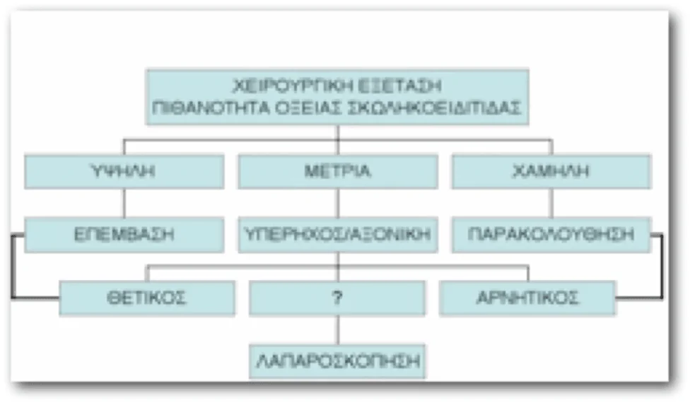
Laboratory and radiological tests in acute appendicitis
White blood cells are mildly elevated in typical appendicitis but may be completely normal (<10,000) in 10% of patients. In the disease complicated by peritonitis, on the contrary, they are greatly increased >20,000. The inflammatory marker CRP is also elevated, but not always.
Apart from blood tests, the contribution of ultrasound to the differential diagnosis of the disease is important. Due to its convenience and non-radiation, ultrasound is the examination of choice to confirm acute appendicitis and at the same time rule out other diseases in the area, especially in women and children. In this area, we often find gynecological diseases from the right ovary and fallopian tube that mimic appendicitis, such as ruptured ovarian cyst, pyofallopian tube.
Computed tomography can also be used in adults mainly for cases where the clinical picture is indeterminate and complicates the diagnosis. By using all these tests we have succeeded in reducing unnecessary appendectomies in recent years to 10-15%.
Nevertheless, because appendicectomy as an operation has clearly fewer complications and mortality than ruptured appendix and peritonitis, surgery is definitely preferable to waiting.
In this rare case where we cannot establish the diagnosis with certainty, especially when it comes to a female, the appropriate approach is diagnostic laparoscopy. During laparoscopy, the laparoscope camera is inserted through a small 1.5 cm incision in the patient's abdomen and all internal organs are checked under direct vision. Once the existence of acute appendicitis is established, two other laparoscopic tools are placed through corresponding small incisions and the operation is completed laparoscopically.
Appendicitis Differential Diagnosis
There are certain conditions that resemble or are confused with acute appendicitis. In preschool children, intussusception (two adjacent parts of the intestine enter each other), Meckel's diverticulum, acute gastroenteritis can mimic the disease. In older children, the diagnosis should also consider mesenteric lymphadenitis (it is a microbial infection of the area characterized by swelling of the lymph nodes on ultrasound), as well as inflammatory bowel diseases (ulcerative colitis, Crohn's disease).
In adults, conditions of the area include pyelonephritis (infection of the kidney), colitis, diverticulitis, pyosalpingus, ruptured ovarian cyst, ovarian torsion, ectopic pregnancy, endometriosis, and more.
The occurrence of acute appendicitis during pregnancy is also a serious problem. The diagnosis is often delayed as the pregnancy itself causes nausea, anorexia, an increase in white blood cells and only the pain (which, however, is not typical as after the 5th Month the uterus presses on the intestine and the appendix rises high away from its classic position) can make us suspicious. When ultrasound cannot help in some cases MRI is recommended (CT is prohibited due to radiation). The pregnant woman must be operated on immediately because the miscarriage rates reach 10% if peritonitis occurs.
Another category of patients is the elderly, where the symptoms are often mild and the diagnosis may be delayed. The lack of symptoms in almost half of the elderly patients is the main reason for delayed diagnosis and an increase in the peritonitis rate of more than 50% in these ages compared to less than 20% in the young. Other conditions confused with acute appendicitis in the elderly are tumors, ulcer perforation, diverticular rupture, acute cholecystitis, intestinal torsion, intestinal ischemia or thrombosis, and others. Computed tomography is the ideal test for these patients.
Treatment of Appendicitis
The treatment of acute appendicitis is its surgical excision. Antibiotic use that covers both aerobic and anaerobic microbes found in the gut is indicated before and after surgery. Hospital stay in uncomplicated cases is 1-2 days. If peritonitis occurs, hospital stay and antibiotic administration are prolonged until the patient is afebrile.
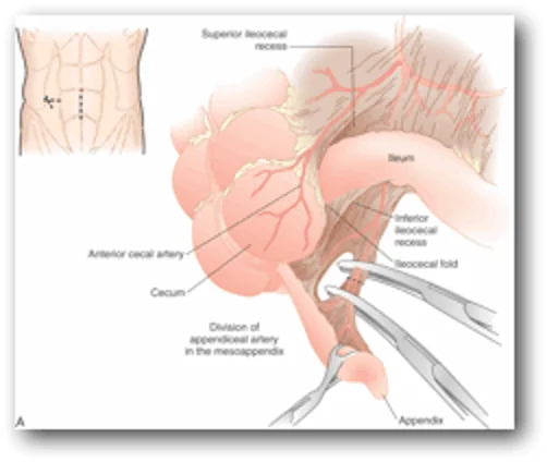
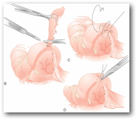
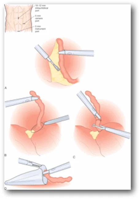
The operation can be done open or laparoscopically. The results of both are equally good, but the laparoscopic technique is preferred in women (to rule out possible gynecological diseases), in the obese (technically easier and with less morbidity), in the elderly and in cases where there is doubt about the diagnosis. In recent years there has been a tendency to increase laparoscopic removal despite its increased cost.
Appendiceal abscess is a complication of acute appendicitis that was not treated in time. The patient presents with a mass in the appendix area and fever. This case is initially treated with antibiotics and drainage of the abscess under ultrasound or axial, and in the second year surgery follows.
Another category of patients is that of chronic (recurrent) appendicitis. These are repeated episodes of pain in the appendix area, but without the classic clinical picture. The remaining findings in the hematological and radiological tests may be indicative of the condition, so the treatment is the removal of the appendix. But there are also cases where everything looks normal. These cases tend to be characterized as chronic abdominal pain and the cause is rarely the appendix, and its removal does not improve the symptoms. These patients need further screening with a colonoscopy and other tests to find the cause of the pain.
An interesting question that the surgeon may face is how much he should remove an apparently normal appendix in a patient who went to surgery because of signs of acute appendicitis. The answer is rather affirmative, the appendix that looks normal during the operation is better to be removed because the complication rates are very small and often other diseases of the appendix (malignancy, Crohn's disease, etc.) are found in the biopsy. However, in addition to its removal, an extensive laparoscopy of the abdomen should also be performed in order to find any other cause of the symptoms and pain.
Finally, regarding the complications of the operation, these are mainly the infection of the incision in a percentage of 15-20% when there is a rupture of the appendix, the ileus (1-3%), while the fertility in women seems not to be affected.
Appendicitis Conclusions
Acute appendicitis is one of the most common surgical emergencies. We should immediately contact a surgeon when we have persistent severe pain in the abdomen, especially in the lower right. Early surgical treatment ensures minimal complications from the disease, but if neglected, it can have unpleasant consequences for the patient.