
Gall laparoscopy – Stones polyps symptoms treatment.
Cholelithiasis cholecystitis stones mud polyps.
The bile is a common medical problem whether it is due to mud or stones, or a polyp. OR laparoscopic cholecystectomy (removal of bile laparoscopically through small holes) is today one of the most frequent surgical operations. The operation is performed through 4 small holes (5-10 mm) in the abdomen, compared to a large incision of 7-10 cm that was made with the old method that is now applied in a few exceptions. The advantages are minimal postoperative pain and faster recovery and return to daily activities. Hospitalization usually lasts 24 hours.
Gallbladder – Bile
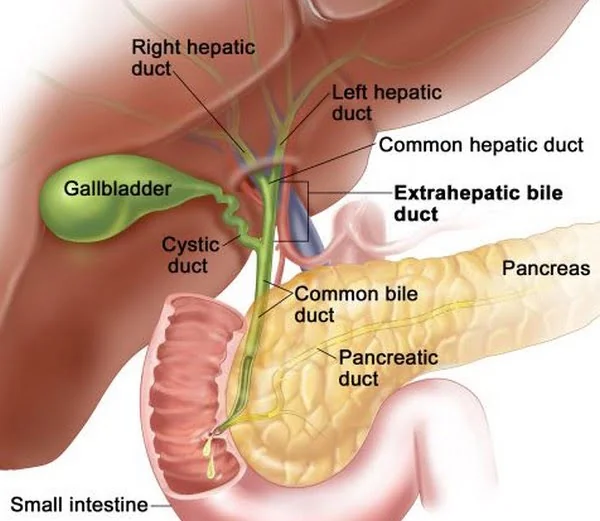
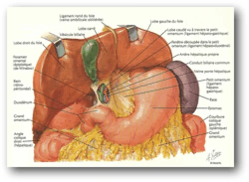
The gallbladder is a sac-shaped organ located under the right side of the liver.
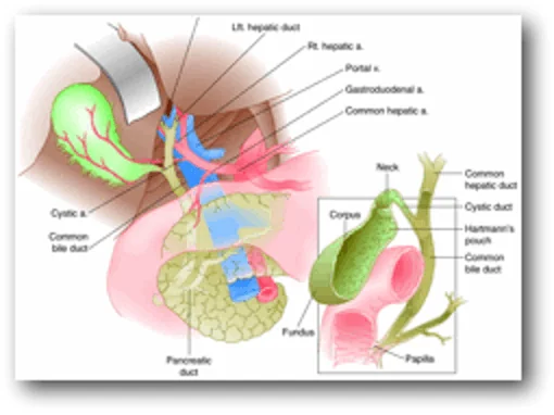
It stores the bile produced by the liver and which participates in the digestion of fats and fat-soluble vitamins. Bile is released from the gallbladder after eating and is transported through small and thin tubes into the duodenum (small intestine).
The majority of patients do not experience any digestive disorders after gallbladder removal.
Gall pathology.
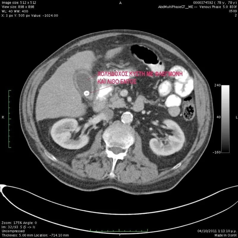
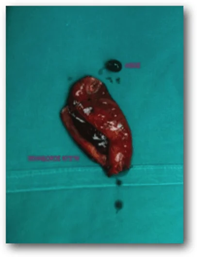
The problems are usually caused by the presence of gallstones in the gallbladder. Gallstones (gallstones) as their name describes are small hard "stones" formed from cholesterol and bile salts in the gallbladder or bile duct.
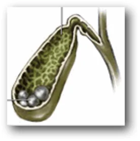
Why stones begin to form in an otherwise normal gallbladder is not known. However, the mechanism by which gallstones are created has to do with the change in the ratio of the components of bile.
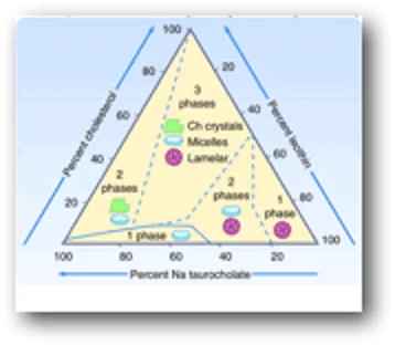
Unfortunately, there is no way to prevent the formation of gallstones.
The complications of gallstones are as follows:
A) To prevent the flow of bile out of the gallbladder, resulting in its swelling. This causes acute abdominal pain (colic), vomiting, dyspeptic complaints.
B) If inflammation is caused in addition, then the pain becomes continuous, fever appears and then we talk about acute cholecystitis.
C) In the event that the stone (gallstone) moves towards the tube from where the bile enters the intestine (bile duct) then jaundice can be caused (yellowing of the skin, eyes, dark urine, white stools),
D) In some cases even pancreatitis.
Bile diagnosis and treatment.
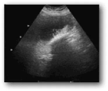
Diagnosis begins with clinical examination and history. The most common symptoms of gallstones are colicky pain in the right upper quadrant of the abdomen often associated with eating, dyspeptic complaints, nausea. When inflammation develops the pain becomes continuous, intense, fever appears and there is sensitivity in the area.
Ultrasound is the test of choice used to highlight gallstones and inflammation and is usually combined with blood tests (WBC, CRP, BIL, GGT, SGOT, SGPT etc).
In complicated cases, additional tests may be needed to establish the diagnosis.
Cholelithiasis does not improve over time, on the contrary, it worsens, and therefore conservative and pharmaceutical measures are only offered as a temporary solution until the gallbladder is removed.
Laparoscopic removal of the gallbladder is the operation of choice and the safest treatment for gallstones and cholecystitis.
Bile preparation for surgery.
Upon your admission to the hospital, the pre-operative check-up takes place. You should not eat or drink for at least 6 hours before the operation. You must have bathed before surgery and if you have a problem with constipation an enema is recommended. If you are taking medication you must inform your surgeon who will tell you if you need to stop certain of them (be especially careful with aspirin, anticoagulants or anti-inflammatory drugs). The doctor may also ask you for a unit of blood.
Laparoscopic Gallbladder Surgery Method Technique
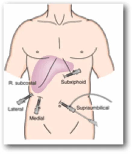
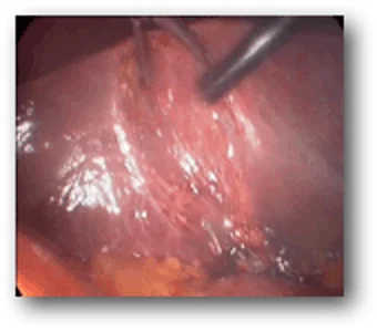
The operation is performed under general anesthesia. Through a small incision in the navel, the camera (laparoscope) is inserted which transfers the image of the inside of the abdomen to a screen. The abdominal cavity is filled with gas to inflate and provide good visibility and space for tools to move. Through three other incisions, laparoscopic instruments are inserted which allow the surgeon to detach the gallbladder from the liver.
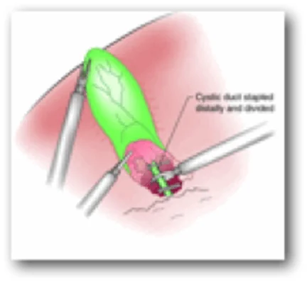
If there are stones in the bile duct, they may be removed first by the Gastroenterologist with ERCP followed by surgery, or they may be removed laparoscopically along with the gallbladder using a choledochoscope.
Open cholecystectomy.
In a small number of patients the laparoscopic method is not possible or not indicated.
The main reasons are:
- Suspicion of malignancy
- Other concomitant diseases that prohibit laparoscopy (respiratory, cardiovascular, coagulation disorders, generalized peritonitis, ileus)
- Stone size
- Chronic inflammation
- Abnormality of the anatomy of the area
- Previous operations
- Reasons for conversion from laparoscopic to open surgery: Bleeding Poor visibility Bile duct injury Any doubt about completing the procedure laparoscopically
General information about laparoscopic cholecystectomy.
The hospital stay is usually 24 hours. Returning to work is possible after a few days depending on the type of work. Conversely, patients who undergo open surgery usually return to work after 3-4 weeks. The scars from the incisions are very small 5-10 mm, and the risk of hernia is reduced. Complications from a laparoscopic cholecystectomy when performed by a properly trained and experienced surgeon are comparable or less than the open method. The onset of fever, jaundice (yellowing of the skin or eyes), increasing abdominal pain, vomiting are possible indications that there is a complication and your surgeon should be informed.
Bile special cases.
The gall bladder also presents other conditions which are quite rare such as cancer and movement disorders. The motor dysfunction of the gallbladder, which has begun to be diagnosed in recent years in patients with biliary-type pain, but without findings of cholelithiasis or cholecystitis and with a normal upper and lower digestive examination (gastroscopy, colonoscopy, CT, MRCP), is a difficult diagnostic problem. The dysfunction of the sphincter of Oddi is included in the context of motor disorders. These conditions often coexist with irritable bowel syndrome (spastic colitis), diabetes, hyperlipidemia, obesity, scleroderma, etc.